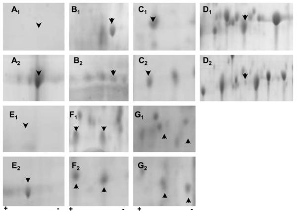
Fig. 3.
Posttranslational modifications evident in ARPE19 monolayers exposed to either unmodified matrix (BM) (a1-g1) or glycation (AGE-BM) (a2-g2). The most significantly altered 2D regions are highlighted for protein alterations occurring with and without matrix modification. Arrowheads indicate proteins of interest correlating with a selection of data in Table 1. Identified proteins include a: α enolase phospho D glycerate (47 kDa); b: vimentin (53.5 kDa); c: nucleoside diphosphate kinase A (17.1 kDa); d: heat shock protein beta1 (22.8 kDa); e: stress-70 protein (73.6 kDa); f: barrier to auto-integration factor (10 kDa) nuclear transport factor 2 (14.5 kDa); g: peroxiredoxin 2 (21.9 kDa); lactoylglutathione lyase (20.6 kDa)

