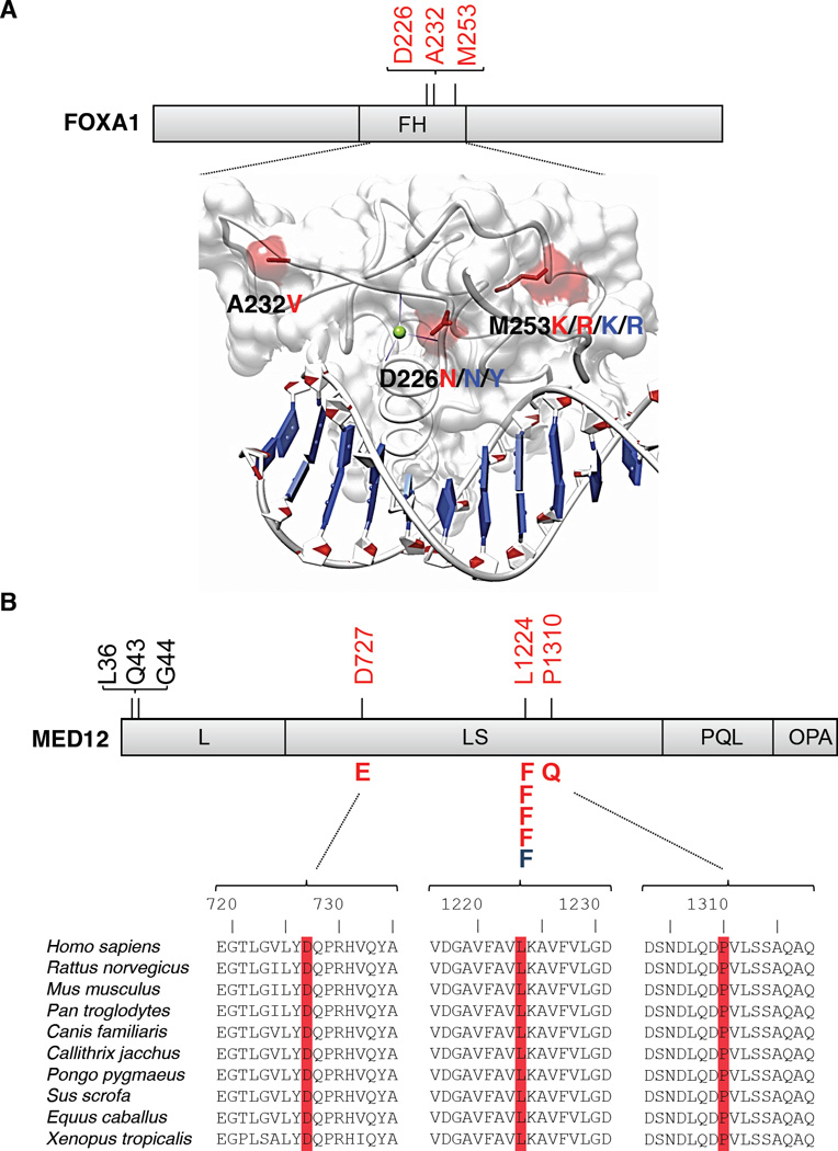
Fig 2. Recurrent somatic mutations in FOXA1 and MED12.

Mutations detected by exome sequencing are depicted (red), as are variants from non-overlapping transcriptome sequencing data (blue). (A) Structural analysis of mutations in FOXA1. Mutated residues are mapped to the structure of the HNF3γ fork-head domain from coordinate file 1VTN.pdb (www.pdb.org)24 and highlighted in red. FH, Fork-head domain. (B) Recurrent MED12 mutations in prostate cancer (red, blue) are distinct from those reported in uterine leiomyeoma (shown in black)28. Domains of MED12 are denoted as in Zhou et al.25. Multispecies conservation of the mutated sites is shown below the mutation.
