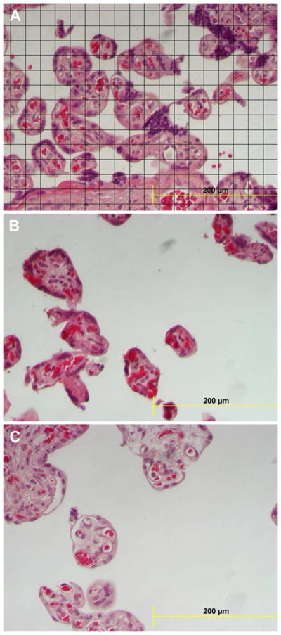
Fig. 1.

(A) Section of villi with the overlying grid used in the morphometric evaluation. (B) Section from a placenta with FGR showing reduced terminal villi and villous surface area per unit area of the field. (C) Section from a placenta of a patient with both preeclampsia (PE) and fetal growth restriction (FGR) showing both reduced terminal villous volume and surface area with a prominent intervillous space.
