
Figure 1.
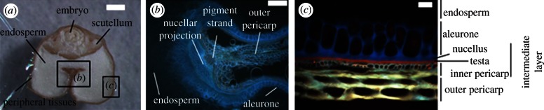
Morphological structures of cross-sectioned wheat grain as seen under the stereomicroscope (a) thick section (scale bar, 500 µm) and under the microscope using a UV-light excitation source (b) thin section (scale bar, 200 µm); (c) thin section (scale bar, 20 µm).