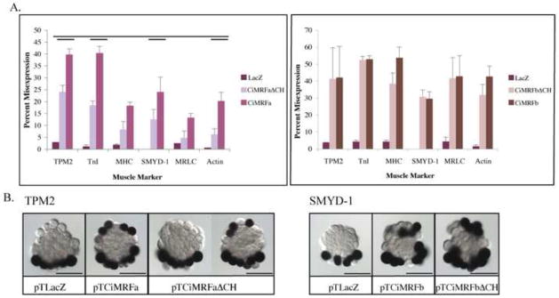
Figure 3. Effect of ΔC/H mutation on myogenicity of Ci-MRF.

Panel A (left): ΔC/H mutant CiMRFa drove the expression of all six muscle markers in the notochord at levels significantly lower than wild type CiMRFa (single bar denotes p<0.05, double bar denotes p<0.01). Error bars represent standard deviation. Panel A (right): ΔC/H mutant transcripts of CiMRFb drove the expression of all six muscle markers in the notochord in embryos at levels that were not significantly different from embryos electroporated with wild type CiMRFb. Error bars represent standard deviation. Panel B (left): In situ hybridization showing TPM2 expression in embryos electroporated with the designated plasmid. Panel B (right): In situ hybridization showing SMYD-1 expression in embryos electroporated with the designated plasmid. Scale bars are 50μm.
