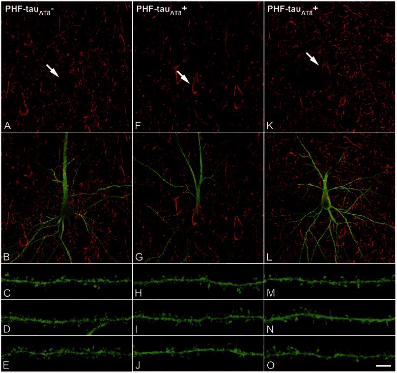
Figure 5.
PHF-tau− and neurons with Pattern IIa PHF-tauAT8 staining. Neurons and dendrites in the CA1 of Patients P14 (A–J) and P13 (K–O), injected with Lucifer yellow, and with a soma free of PHF-tauAT8 ( ; A–E) or with PHF-tauAT8–ir tangles (Pattern IIa; F–O). (A–B, F–G and K–L) Stacks of 18 and 35 images, respectively, obtained after combining the channels acquired separately for Lucifer yellow (green) and PHF-tauAT8 (red). (C–D, H–J and M–O) Stacks of 26–58 optical confocal sections of the basal dendrites of
  (A and B) and immunostained (
 ; F–G, K–L) Lucifer yellow-injected pyramidal neurons. These
  and
and  Lucifer yellow-injected neurons were recovered and reprocessed immunocytochemically using anti-PHF-tauPHF-1. The
  neuron was non-immunostained for PHF-tauPHF-1 (PHF-tau−), whereas the
  neurons were also PHF-tauPHF-1 (Supplementary Fig. 5). Scale bars: A, F and K = 10 µm; C–E, H–J and M–O = 3 µm.

