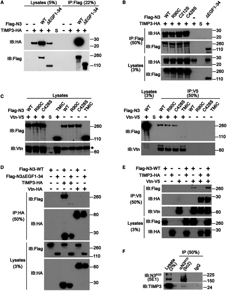
Figure 4.
TIMP3 forms complexes with Notch3ECD and Vitronectin. (A–D) FLAG-tagged wild-type NOTCH3 (Flag-N3-WT) and mutant NOTCH3 (ΔEGF1-34, R90C, C212S, C428S, TMIC) were cotransfected with haemagglutinin-tagged TIMP3 (TIMP3-HA) (A and B) or V5-tagged Vitronectin (Vtn-V5) (C) in 293T cells. Forty-eight hours after transfection cells were harvested and immunoprecipitated (IP) with anti-FLAG antibody (A and B) or anti-V5 antibody (C) and subjected to immunoblot (IB) with the indicated antibodies. (D) FLAG-tagged wild-type NOTCH3 (Flag-N3-WT) and FLAG-tagged NOTCH3 deletion mutant lacking the 34 EGF-like repeats (Flag-N3ΔEGF1-34) were co-transfected with TIMP3-HA or haemagglutinin-tagged VTN (Vtn-HA). Immunoprecipitation was performed with anti-haemagglutinin (HA) antibody. (E) Flag-N3-WT was co-transfected with TIMP3-HA and Vtn-V5 and IP was performed with anti-V5 antibody. This panel is representative of three independent experiments. (F) Interaction between endogenous NOTCH3 and TIMP3 in coronary artery smooth muscle cells. NOTCH3 immunoprecipitation was performed using anti-Notch3ECD rabbit polyclonal antibody (BC2), normal rabbit IgGs were used as controls. Asterisk indicates non-specific labelling. A representative gel of at least two independent experiments is shown.

