Figure 5.

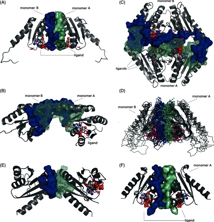
Dimerization pattern of USP family members. (A) probable dimer assembly of USP NE1028 from N. europea [Protein Data Bank (PDB) code: 2PFS]; (B) likely incorrect dimeric assembly of USP NE1028 from N. europea (PDB code: 2PFS) predicted by the PISA server; (C) dimeric assembly of UspE protein Rv2623 from Mycobacterium tuberculosis (PDB code: 3CIS); (D) superposition of type 1 dimers (representatives listed in the Table 1); (E) Incorrect UspF assembly (PISA AB); (F) Correct assembly (PISA AA) of UspF (PDB code: 3FDX) ATP-binding residues are shown in pink, dimerization interface residues from monomers A and B are shown in green and blue respectively, and ligand molecules are shown in CPK colors in either space-filling or ball-and-stick representation.
