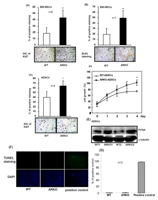
Figure 4. Depletion of AR in BM-MSCs and ADSCs stimulates their proliferation.
(A) IHC staining result of Ki67. BM-MSCs isolated from 8 weeks old WT and ARKO mice were seeded on culture plates. Percentage of positive staining was expressed as positive Ki67 signals/total nuclear signals, n = 6. (B) BrdU labeling experiment result. Percentage of positive staining was calculated as positive BrdU signals/total nuclear signals, n = 7. (C) IHC staining result of Ki67 in ADSCs that were isolated from 8 weeks old WT and ARKO mice, n = 4. (D) MTT assay result of ADSCs of WT and ARKO mice. Percentage of growth was normalized to the highest value (set as 100%). (E) Western blot analysis showing expression of PCNA in ADSCs obtained from the WT (WT1 and WT2) and the ARKO (ARKO1 and ARKO2) mice. α-Tubulin was used as control. (F) TUNEL staining was performed according the manufacturer’s instruction to examine apoptotic death of BM-MSCs of WT and ARKO mice. Positive control provided in the TUNEL assay kit was applied. Green fluorescence signal represents the positive apoptosis signal. (G) Quantitation of TUNEL staining results. *, p-value < 0.05, **, p-value < 0.01, when compared to controls.

