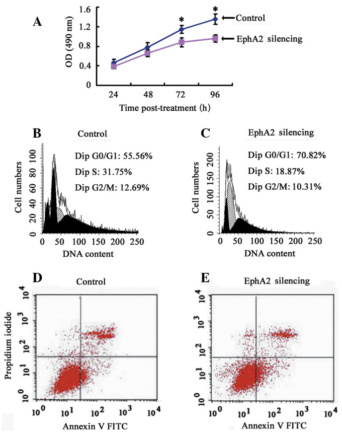
Figure 3.

The effect of EphA2 on the proliferation, cell cycle distribution and apoptosis of NPC 5-8F cells. (A) Cell proliferation was measured by the CCK-8 assay every 24 h for 4 days. (B and C) Differently treated cells were stained with propidium iodide and analyzed by flow cytometry. Proportion of cells in various phases of the cell cycle. The results are the means of three independent experiments ± SD (P<0.05). (D and E) Cells staining positive for Annexin V-FITC and negative for PI were considered to have undergone apoptosis. Average apoptotic rate of three independent experiments ± SD are shown. *P<0.05.
