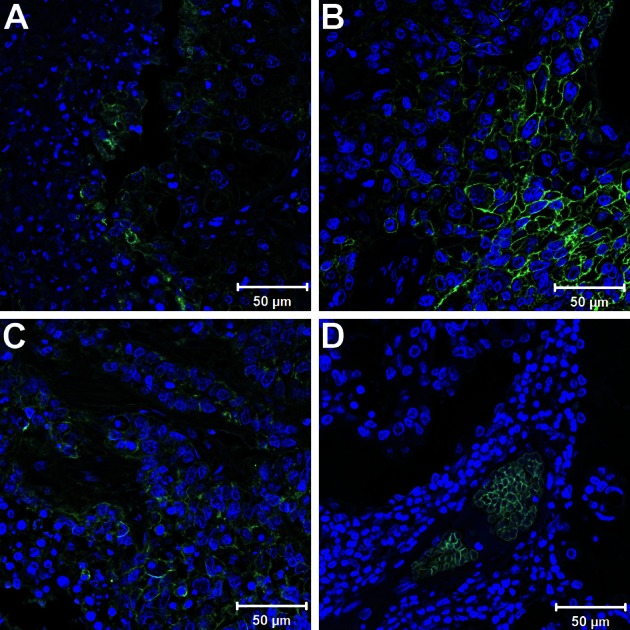
Figure 3.

Glucose transporter-1 (Glut-1) staining of different lung neoplasms. A-C, Immunofluorescence staining of paraffin-embedded sections shown as merged image of Glut-1 (green) and 4,6-diamidino-2-phenylindole-2-HCl (blue) demonstrates positive surface Glut-1 lung tumor staining in adenosquamous carcinoma (A), squamous cell carcinoma (B), and adenocarcinoma (C). D, In contrast, sections from bronchoalveolar carcinoma show Glut-1 staining in erythrocytes, but not in malignant cells.
