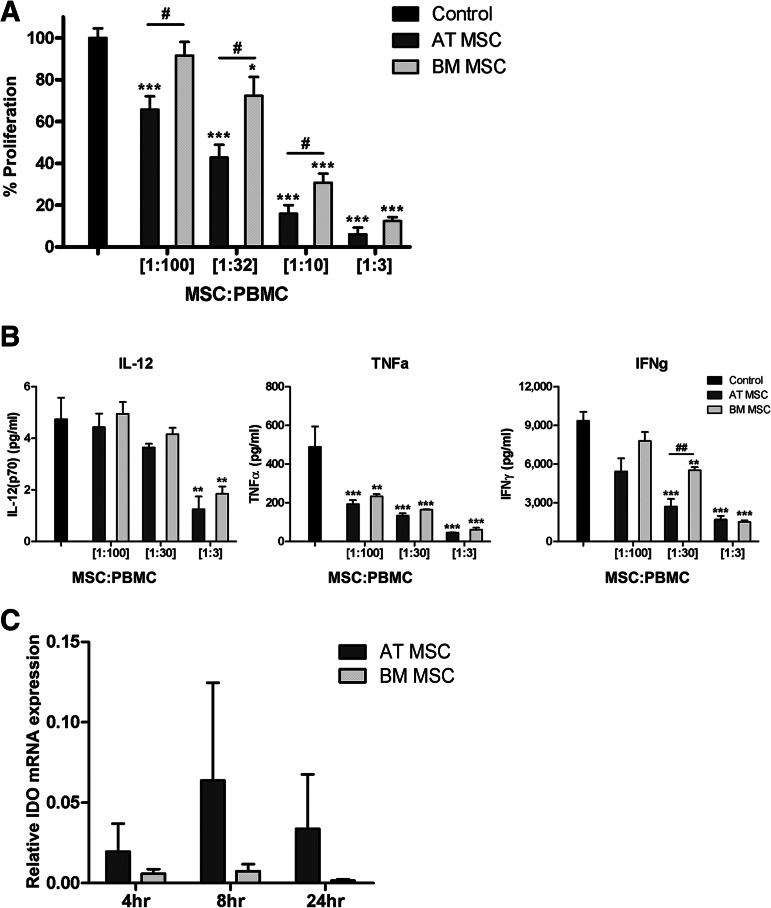
Figure 3.
AT-MSCs are more potent in suppressing PBMC proliferation compared with BM-MSCs. (A): MSCs suppressed PBMC proliferation in a dose-dependent fashion. AT-MSCs showed a significantly stronger suppression of proliferation at MSC:PBMC ratios of 1:100, 1:32, and 1:10 (two separate experiments; n = 9 for AT-MSCs, n = 8 for BM-MSCs). (B): From one experiment, culture supernatants of PBMC proliferation in the presence and absence of MSCs were assayed for cytokine concentrations at day 5 of coculture (n = 3 for both groups). Statistical analysis was performed using Student's t test (data are mean ± SEM; * indicates compared with control: *, p < .05; **, p < .01; ***, p < .001; # indicates AT-MSCs compared with BM-MSCs: #, p < .05; ##, p < .01). (C): After IFN-γ stimulation of MSCs, both BM-MSCs and AT-MSCs showed IDO mRNA upregulation, with an optimum at 8 hours. IDO mRNA expression is shown relative to β-actin mRNA expression. Statistical analysis was performed using Student's t test (n = 3 for both groups). Abbreviations: AT MSC, adipose tissue-derived multipotent stromal cells; BM MSC, bone marrow-derived multipotent stromal cells; IDO, indoleamine 2,3-dioxygenase; MSC, multipotent stromal cells; PBMC, peripheral blood mononuclear cells.

