Figure 1.
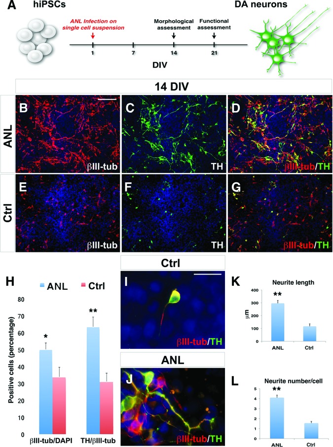
Differentiation of IMR90-hiPSCs into DA neurons after ANL overexpression. (A): Schematic representation of the one-step differentiation protocol. DA neurons are characterized after 14 DIV from viral transduction. (B–G): Immunocytochemical analysis for TH and βIII-tubulin in ANL-infected (B–D) and Ctrl (E–G) IMR90-hiPSC-derived neurons. (H): Quantification of TH+/βIII-tubulin+ and βIII-tubulin+/4′,6-diamidino-2-phenylindole (DAPI) yield. (I, J): High magnification of βIII-tubulin/TH immunostaining highlights a more differentiated morphology in IMR90-hiPSC-derived neurons infected with ANL (J) when compared with control (I). (K, L): Comparison of neurite length and number between ANL and control conditions. The nuclei are stained with DAPI. Scale bars = 100 μm (B–G) and 20 μm (I, J). Student's t test was used. *, p < .05; **, p < .01. Abbreviations: ANL, ASCL1, NURR1, and LMX1A; Ctrl, control; DA, dopamine; DIV, days in vitro; hiPSCs, human induced pluripotent stem cells; TH, tyrosine hydroxylase; βIII-tub, βIII-tubulin.

