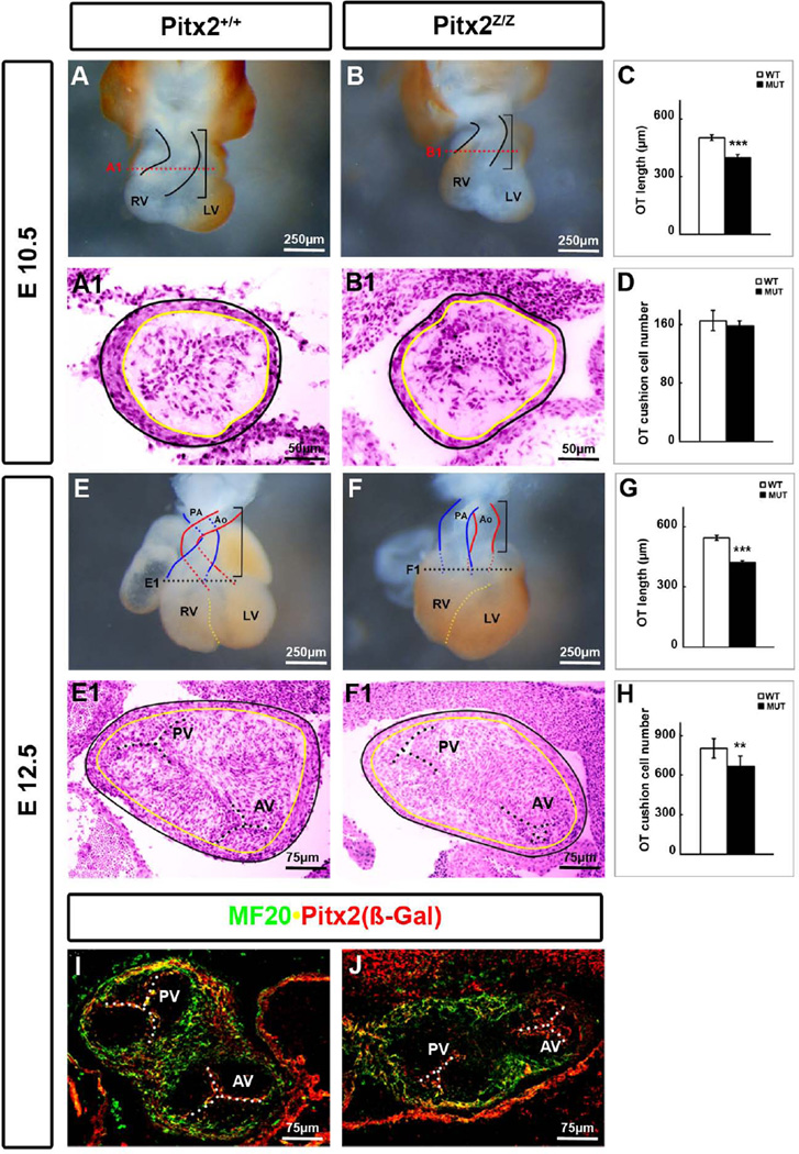
Figure 1. Shorter and Hypocellular Cardiac OT in Pitx2 Mutants.
Ventral view of the entire heart at E10.5 (A, B) and E12.5 (E, F) showed a shortened OT with a prominent DORV in Pitx2-mutant mice (F). (C, G) The length of the OT was measured as indicated by brackets. Statistics were based on results from 3 different embryos at each stage. HE staining on 14 µm transverse cryosections at E10.5 (A1, B1) and E 12.5 (E1, F1) mice indicated thinner OT epithelium in the conus. The black and yellow lines correspond to the outer and inner epithelium, respectively. (D, H) Cell counts of a set of 5–8 serial sections along the OT showed reduction of cells in the cushions of mutants. ***: p<0.01, **: p<0.05. (I, J) Double labeling immunohistochemistry on E12.5 mouse transverse sections for MF20 and Pitx2 (β-Gal). No MF20+ cells were detected in the conal septum and semilunar valves in the mutants. Ao, aorta; AV, aortic valve; LV, left ventricle; PA, pulmonary artery; PV, pulmonary valve; RV, right ventricle.

