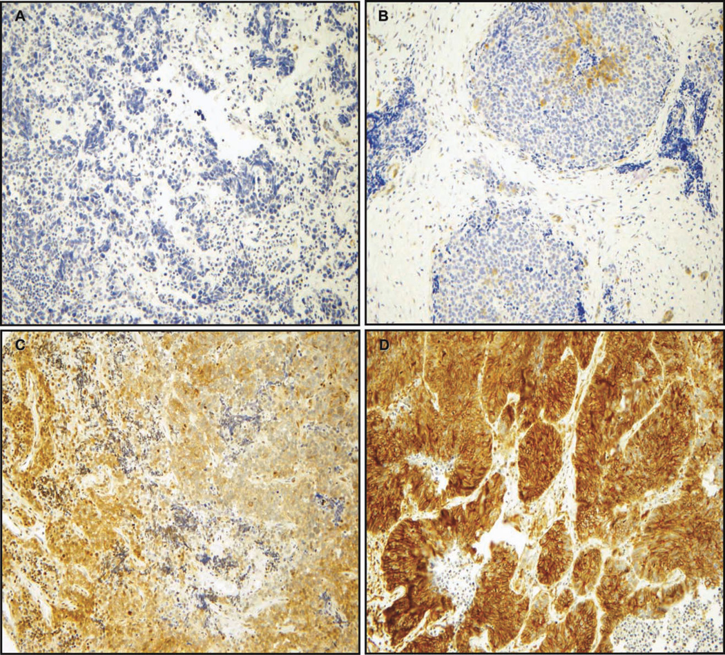
Figure 2.
Phospho-paxillin Tyr118 expression was evaluated via immunohistochemistry. A tissue microarray consisting of SCLC tissue samples from 35 patients was incubated with an antibody against phospho-paxillin Tyr118. Phospho-paxillin Tyr118 displayed a cytoplasmic staining. The staining intensity of the SCLC cases was evaluated by 2 independent observers (A.L.G., S.O.) using the following scoring system: 0, no staining; 1, weak; 2, moderate; and 3, strong. The staining intensity was then multiplied by stained tumor cell percentage to obtain the final staining score (range, 0–300). Representative images are shown for SCLC tumors with (A) absent, (B) weak, (C) moderate, and (D) strong phospho-paxillin expression. (Original magnification, ×200.)

