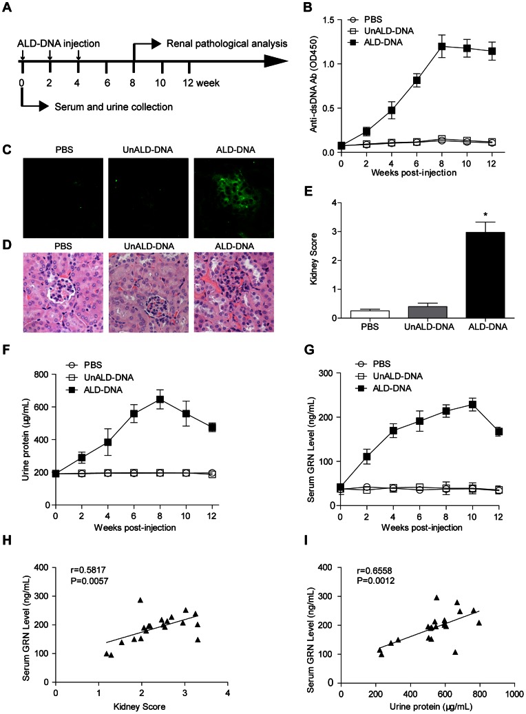
Figure 1. Serum granulin (GRN) levels were up-regulated in lupus model and were associated with the severity of lupus nephritis.
(A) Schematic diagram of DNA injection. 6- to 8-week old female BALB/c mice were injected subcutaneously with ALD-DNA (50 µg/mice) plus CFA at week 0, followed by two booster injections of ALD-DNA (50 µg/mice) emulsified with IFA at week 2 and week 4 after initial injection. (B) Serum anti-dsDNA IgG levels were measured by ELISA every 2 weeks after initial injection. Data are means ± SD from 8 mice in each group. (C) 8 weeks after initial injection, glomerular immune deposition were detected by direct immunofluorescence for IgG in frozen kidney section from ALD-DNA-injected SLE mice or control mice. Representative images (magnification×200) of 8 mice are shown for each group. (D) Nephritic pathology was evaluated by H&E staining of renal tissues. Images (magnification×200) are representative of 8 mice in each group. (E) The kidney score was assessed using paraffin sections stained with H&E in (D). n = 8. (F) Urine protein levels of the mice were assessed by BCA Protein Assay Kit every 2 weeks. Data are means ± SD from 8 mice in each group. (G) Serum GRN levels were measured by ELISA every 2 weeks after initial injection. Data are means ± SD from 8 mice in each group. (H) The correlation between serum GRN level and kidney score in lupus model. Correlation analysis was performed by Pearson correlation analysis. Each symbol indicates an individual mouse (n = 21). (I) The correlation between serum GRN level and urine protein level in lupus model. Pearson correlation analysis was used to carry out the correlation study. Each symbol indicates an individual mouse (n = 21). *, P<0.05.

