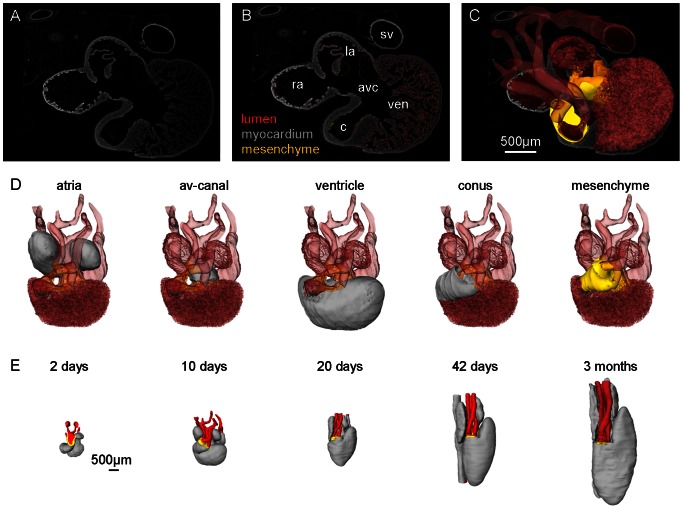
Figure 2. Generation of 3D reconstructions.
A. Example of a 10 µm section from P. guttatus 10 days stained for myocardium with antibodies against rabbit cardiac troponin I. B. Same section as in A with annotations made in Amira®. C. 3D reconstruction based on 160 sections projected out of the section of A with mesenchyme and transparent lumen (myocardium not shown). D. Same 3D reconstruction as in C visualizing the annotated compartments. E. Five reconstructions exemplifying the transition from the youngest to the oldest stage.

