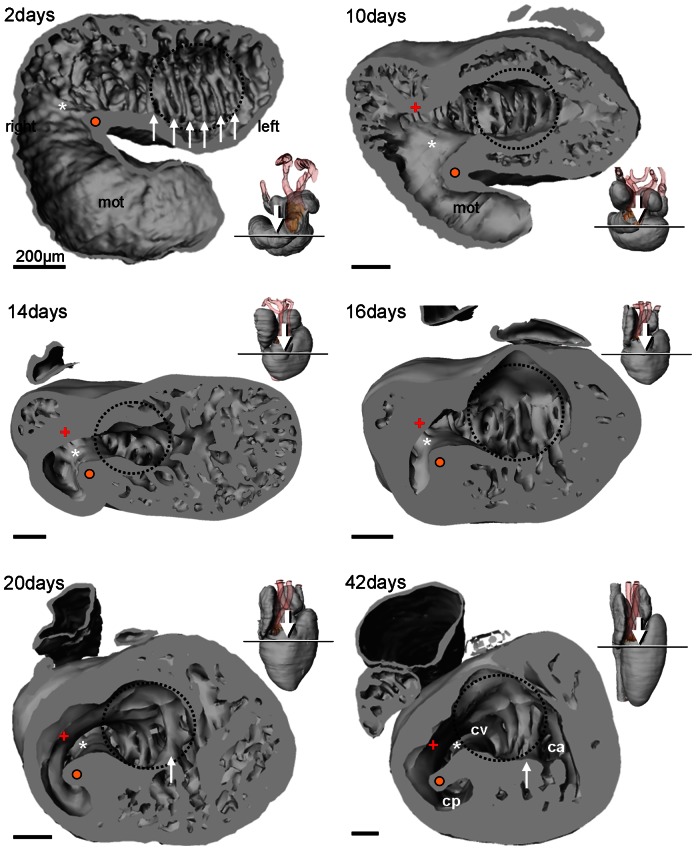
Figure 5. Ventricular morphology of the corn snake.
Looping of the heart tube leaves a fold (white asterisk and orange dot), the bulbo-ventricular fold, on the border of the ventricle and the myocardial outflow tract (mot) (2–10 days). Later, and associated with the ventricularization of the mot, this fold constitutes the muscular ridge (14 days and later), with a free standing part (orange dot) and a complete part (white asterisk). Opposite the muscular ridge, the bulbuslamelle forms (+). The early ventricle has multiple parallel trabecular sheets (white arrows) of equal height but as the ventricle grows (at 20 and 42 days) only one sheet retains a short distance to the atrioventricular canal (indicated by the broken blue line) which may then be annotated as the vertical septum. Inserts show sectioning plane and angle of inspection.

