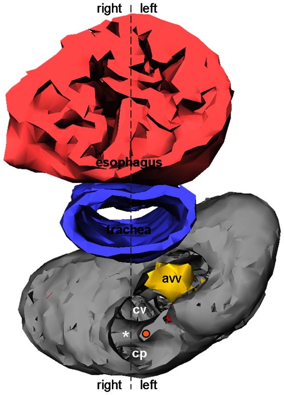
Figure 10. 3D reconstruction of the ventricular base of near-hatching Anolis sagrei (st19).

Using the esophagus and trachea to determine the body midline, it can be seen that the atrioventricular canal remains on the left side. asterisk, complete part of the muscular ridge; avv, atrioventricular valve; cp, cavum pulmonale; cv, cavum venosum; orange dot, free-standing part of the muscular ridge.
