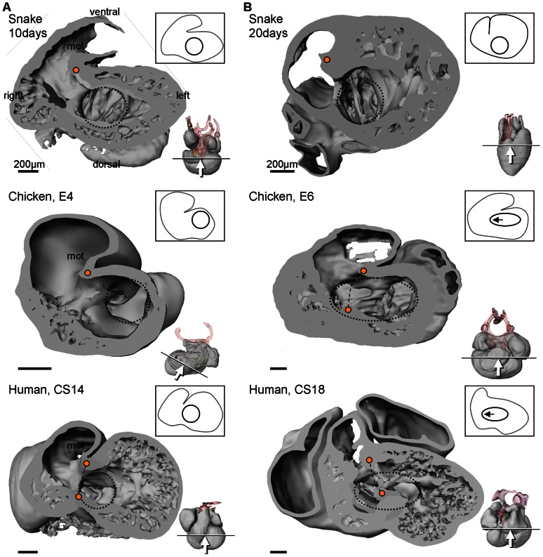
Figure 11. Comparative ventricular development.
A. In early cardiac development, all amniote hearts show a bulbo-ventricular fold (orange dot) on the border of the trabeculated ventricle and the myocardial outflow tract. The atrioventricular canal (blue circle) is exclusively to the left of the fold. B. In non-crocodilian reptiles, the early design is maintained in later development. In amniotes with full ventricular septation (indicated by broken line between two orange dots) the atrioventricular canal expands to the right (note that compared to the stages in A, stages E6 and CS18 are ca. 5% further in gestation, whereas 20 days is ca. 15% further). Miniatures show sectioning plane and angle of inspection. CS, Carnegie stage; E, embryonic day.

