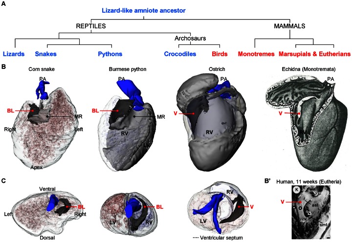
Figure 14. Evolutionary fates of the bulbuslamelle.
A. Phylogenetic tree to show the show the evolution of selected groups of amniotes. B. Ventricles inspected from the right with the ventricular wall made transparent so that the similarity in design between the bulbuslamelle (BL) and the myocardial right atrioventricular valve (V) can be appreciated. The V of birds and monotreme mammals is positioned in the heart where the bulbuslamelle (BL) of the reptile heart is situated. In pythons, the BL is strongly developed and participates in separating the left and right sides of the ventricle. The image of the echidna heart is modified from [70]. B'. Scanning electron microscopic image of the human heart at 11 weeks of development with the wall of the right ventricle removed (modified from [90]). In the human heart, and eutherian mammals in general, the anterosuperior leaflet (asl) of the tricuspid valve is muscular until late in development. C. Cranial view of the ventricular base showing the position of the BL in the ‘basal’ condition (corn snake), in the contributing to separate the left and right sides of the python ventricle and as atrioventricular valve (ostrich) (the ventricle of the corn snake was deformed during fixation, as seen by indentations on the dorsal surface). ap, anterior papillary muscle; smt, septomarginal trabeculation; svc, supraventricular crest; stars, tricuspid gully complex. PA, pulmonary artery.

