Figure 1.

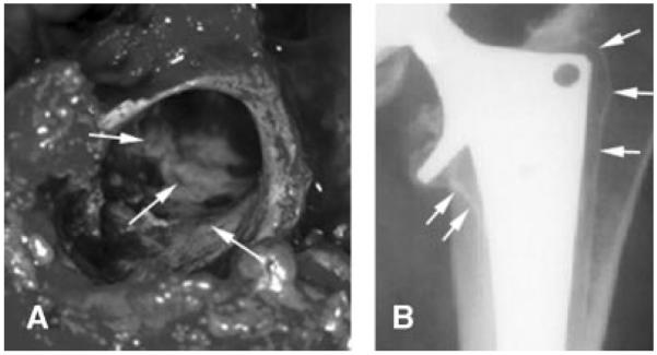
(A) Photograph of the sclerotic bone surface in the proximal femur. (B) Analog preoperative radiograph (anterior–posterior projection) of a loose uncemented femoral component. The sclerotic bone rim is indicated by arrows.

(A) Photograph of the sclerotic bone surface in the proximal femur. (B) Analog preoperative radiograph (anterior–posterior projection) of a loose uncemented femoral component. The sclerotic bone rim is indicated by arrows.