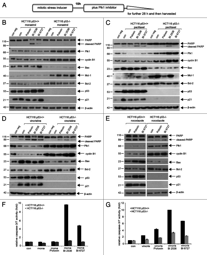
Figure 1. Plk1 inhibitors trigger more apoptosis in HCT116 p53+/+ cells than in HCT116 p53−/− cells in the presence of mitotic stress. (A) Illustration of experimental schedule. Cells were pretreated with a low dose of mitotic stress inducers (15 µM monastrol, 7 nM paclitaxel, 1.2 nM vincristine or 50 ng/ml nocodazole) for 10 h, then Plk1 inhibitor (25 µM Poloxin, 25 nM BI 2536 or BI 6727) was added for further 28 h. (B) Western blot analysis for cells treated with monastrol and Plk1 inhibitors. HCT116 p53+/+ or HCT116 p53−/− cells were treated as described in (A) and harvested for western blot analysis using indicated antibodies. β-actin served as loading control. (C) Western blot analysis for cells treated with paclitaxel and Plk1 inhibitors. (D) Western blot analysis for cells treated with vincristine and Plk1 inhibitors. (E) Western blot analysis for cells treated with nocodazole and Plk1 inhibitors. (F) Quantification of relative activity of caspase-3/7 for cells treated as in (B). (G) Quantification of relative activity of caspase-3/7 for cells treated as in (D). The results are presented as mean ± SD; mona, monastrol; vincris, vincristine.

An official website of the United States government
Here's how you know
Official websites use .gov
A
.gov website belongs to an official
government organization in the United States.
Secure .gov websites use HTTPS
A lock (
) or https:// means you've safely
connected to the .gov website. Share sensitive
information only on official, secure websites.
