
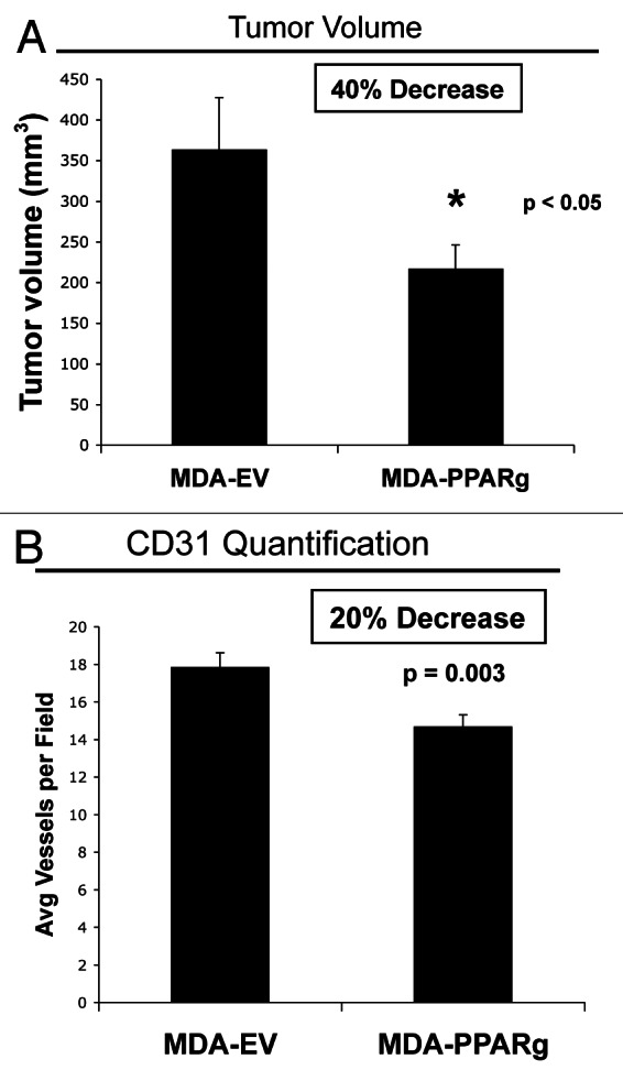
Figure 7. MDA-MB-231 cells overexpressing PPARγ show decreased tumor growth. MDA-MB-231 breast cancer cells harboring EV or PPARγ were injected into the flanks of athymic (immunodeficient) nude mice. (A) Tumor volume is shown after tumor excision 3 weeks post-injection. Note that breast cancer cells overexpressing PPARγ show reduced tumor growth, relative to EV control cells. p values are as shown. n = 10 tumors per experimental group. (B) Tumor angiogenesis. Tumor frozen sections were immunostained with anti-CD31 antibodies. Vascular density quantification (number of CD31-positive vessels per field) indicates that angiogenesis is decreased by 20% in PPARγ tumors.