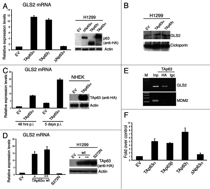
Figure 1. GLS2 expression is regulated by TAp63. (A) H1299 cells were transfected with the indicated HA-tagged p63 constructs and, 24 h after transfection, total RNA was extracted and utilized for reverse transcription and quantitative real-time PCR (qRT-PCR) (left panel) using specific primers for human GLS2 and β actin (for quantity normalization). Results are shown as mean ± SD of three independent experiments. Concomitantly, whole-cell extracts were utilized for western blot analysis using the antibodies to the indicated proteins (right panel). (B) H1299 cells were treated as in (A), and mitochondrial proteins were extracted (see Materials and Methods) and analyzed by IB using antibodies to the indicated proteins. (C) NHEK cells were retrovirally infected with HA-tagged TAp63α expressing virus and 48 h or 5 d after infection GLS2 mRNA levels were measured by q-RT-PCR (left panel). TAP63 protein levels were quantified by immunoblotting (IB) using anti-HA antibody (right panel). (D) H1299 cells were transfected, as indicated, with an empty vector (EV), increasing amount of HA-tagged wild-type TAp63α, or HA-tagged TAp63α (S272N) mutant. Transfected cells were either subjected to RNA isolation for the quantification of GLS2 mRNA by qRT-PCR (left panel) or to immunoblotting for the analysis of the expression levels of wild-type and mutant TAp63α proteins using antibodies to the indicated proteins. (E) Chromatin immunoprecipitation analysis of the human GLS2 promoter was carried out by purifying chromatin from Tet-On/HA-TAp63α-SaOs 2 inducible cell line treated with doxycycline for 24 h and then immunoprecipitating it using HA-specific antibody or IgC-unspecific antibody (see also Materials and Methods). Binding of TAp63α to the MDM2 promoter was used as a control. (F) HEK293E cells were transfected with pGL2 luciferase gene construct holding human GLS2 promoter fragment with either an empty vector (EV) or with the indicated HA-tagged p63 constructs. Co-transfection of a renilla luciferase control plasmid was used to normalize the transfection efficiency. Luciferase assay was performed 24 h after transfection. For all panels, data are shown as the mean ± SD of three replicates.

An official website of the United States government
Here's how you know
Official websites use .gov
A
.gov website belongs to an official
government organization in the United States.
Secure .gov websites use HTTPS
A lock (
) or https:// means you've safely
connected to the .gov website. Share sensitive
information only on official, secure websites.
