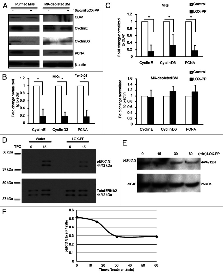
Figure 3. Effect of LOX-PP on ERK1/2 signaling and G1/S phase regulators. (A) BM cells were cultured in StemSpan serum-free media (SFEM) with 25 ng/ml TPO in the presence or absence of 10 µg/ml LOX-PP for 3 d and MKs were purified as described in “Materials and Methods.” The MK depleted BM cells were used for analysis as well. Western blot analysis was performed using antibodies against CD41 (MK marker), Cyclin D3, Cyclin E and PCNA. β-actin was used as a loading control. (B and C) Quantification of western blots was performed using the ImageJ software. Band densities were normalized to β-actin (B) and CD41 (C). Statistical analysis was applied using the Student’s t-test, n = 3, *p < 0.05. (D–F) BM cultures were cultured as in Figure 2A and MKs were isolated using MACS purification (see “Materials and Methods”). MK-enriched BM cultures were then re-cultured in serum-free media for 12 h and treated with vehicle (D) or 10 µg/ml LOX-PP (D and E) for the indicated times. Cell lysates were subjected to western blot analysis using anti-pERK1/2 (D and E) and anti-ERK1/2 (D). eIF4E is used as loading control (E). Shown here is one out of two representative experiments. (F) Quantification of the band density in (E) was performed using the ImageJ software.

An official website of the United States government
Here's how you know
Official websites use .gov
A
.gov website belongs to an official
government organization in the United States.
Secure .gov websites use HTTPS
A lock (
) or https:// means you've safely
connected to the .gov website. Share sensitive
information only on official, secure websites.
