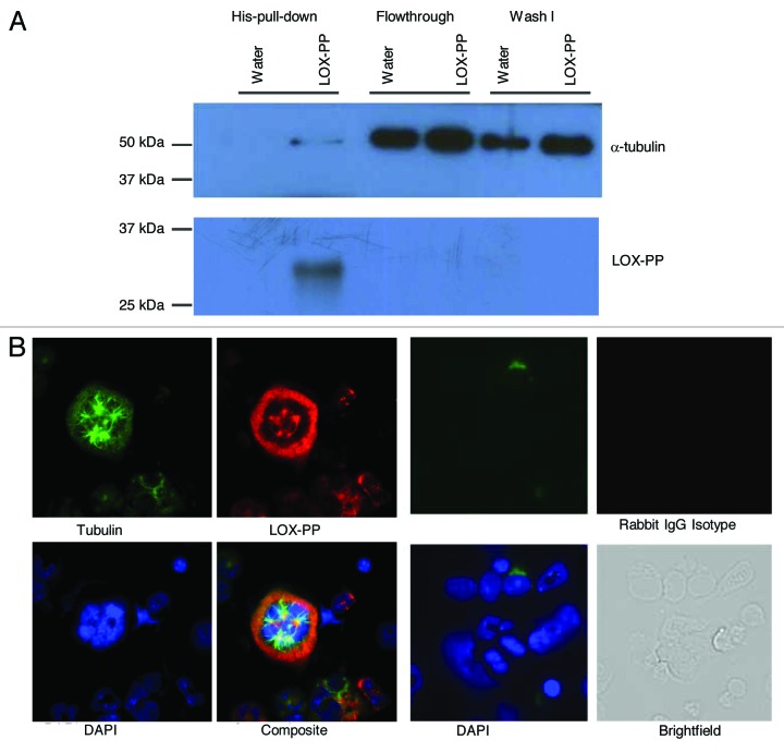
Figure 4. LOX-PP interacts with tubulin in polyploidy MK. (A) Purified MKs were treated with either vehicle (H2O) or 10 µg/ml LOX-PP for 24 h (see “Materials and Methods”). The samples were lysed and incubated with Ni-NTA magnetic agrose beads for 1 h at 4°C. After wash, the elution fractions were examined by western blot analysis with the indicated antibodies. (B) Immunofluorescence analysis of LOX-PP. Mouse BM cells were cultured in the presence of 25 ng/ml TPO in IMDM media supplemented with 10% bovine calf serum BM cells were collected, fixed and stained, as described in “Materials and Methods.” LOX-PP was detected with rabbit polyclonal anti-LOX-PP and Alexa 594 anti-rabbit secondary antibody, α- tubulin was detected with mouse monoclonal anti-tubulin and Alexa 488 anti-mouse secondary antibody, and DNA was stained with DAPI. Staining with rabbit IgG was also performed to verify the specificity of staining with LOX-PP antibody (left panel).

An official website of the United States government
Here's how you know
Official websites use .gov
A
.gov website belongs to an official
government organization in the United States.
Secure .gov websites use HTTPS
A lock (
) or https:// means you've safely
connected to the .gov website. Share sensitive
information only on official, secure websites.
