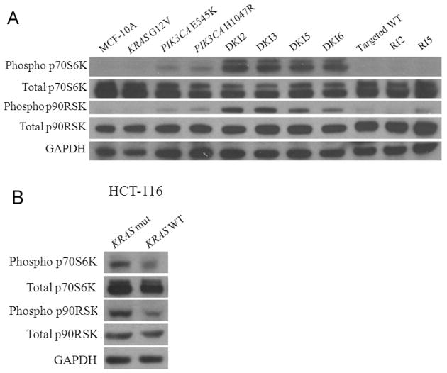
Figure 5. DKI cells have increased phosphorylation of p70S6K and p90RSK.
Western blot demonstrating levels of phosphorylated p70S6K (Thr389), total p70S6K, phosphorylated p90RSK (Ser380), and total p90RSK in cell lines grown in the absence of EGF for A, DKI cells along with MCF-10A and controls,
B, HCT-116 cells with a single copy of mutant KRAS (KRAS mut) and a single copy of wild type KRAS (KRAS WT). GAPDH is shown as a loading control.

