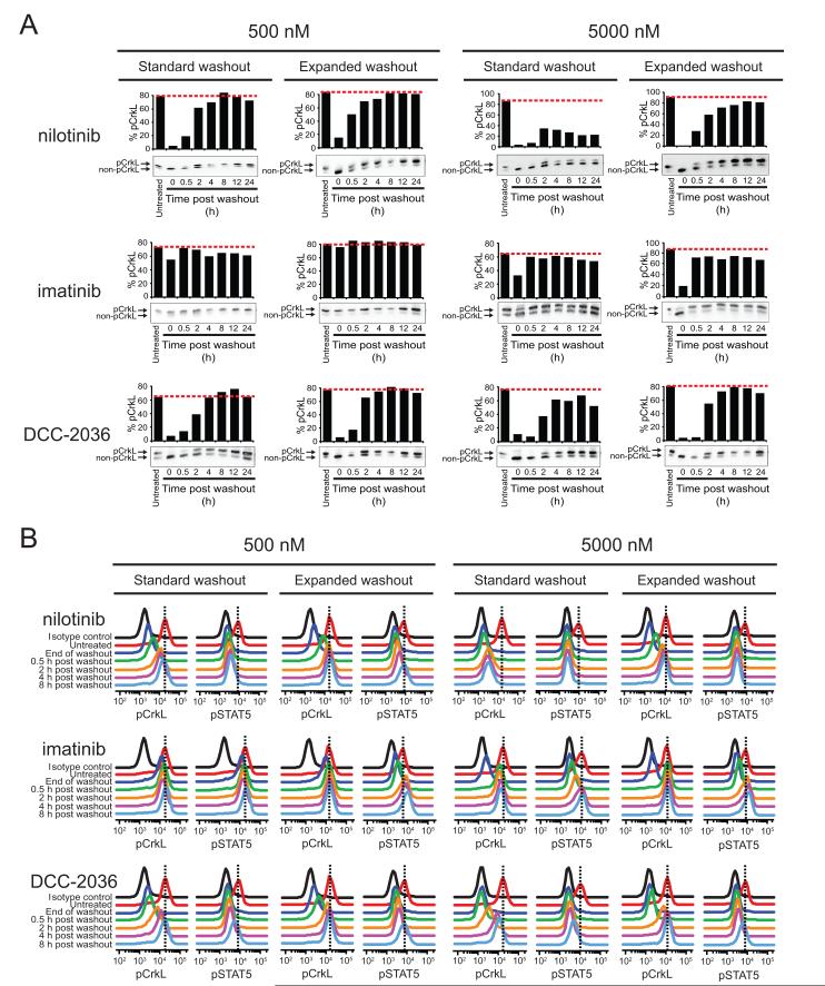
Figure 3. Extent of restoration of BCR-ABL signaling activity following washout of less potent, more selective ABL TKIs tracks with conditions committing cells to apoptosis.
K562 cells were incubated alone or in the presence of 500 and 5000 nM nilotinib, imatinib, or DCC-2036 for 2 h, subjected to standard and expanded washout, and collected at the indicated timepoints post washout for analysis by (A) immunoblot and (B) Phosflow FACS analyses. For immunoblot analyses, the phosphorylated and non-phosphorylated forms of CrkL were resolved by SDS-PAGE, blotted using a total CrkL antibody, and results are expressed as % pCrkL with the red, dashed line indicating the level of % pCrkL in untreated K562 cells. For Phosflow FACS analyses, cells were fixed, permeabilized, and stained using Alexa647-pCrkL and Alexa488-pSTAT5 conjugated antibodies. Results are displayed, for comparison purposes, as the overlaid signal peak traces of isotype control, untreated cells, and each indicated timepoint post washout. Vertical, black dashed lines highlight the peak signal in untreated K562 cells for reference.

