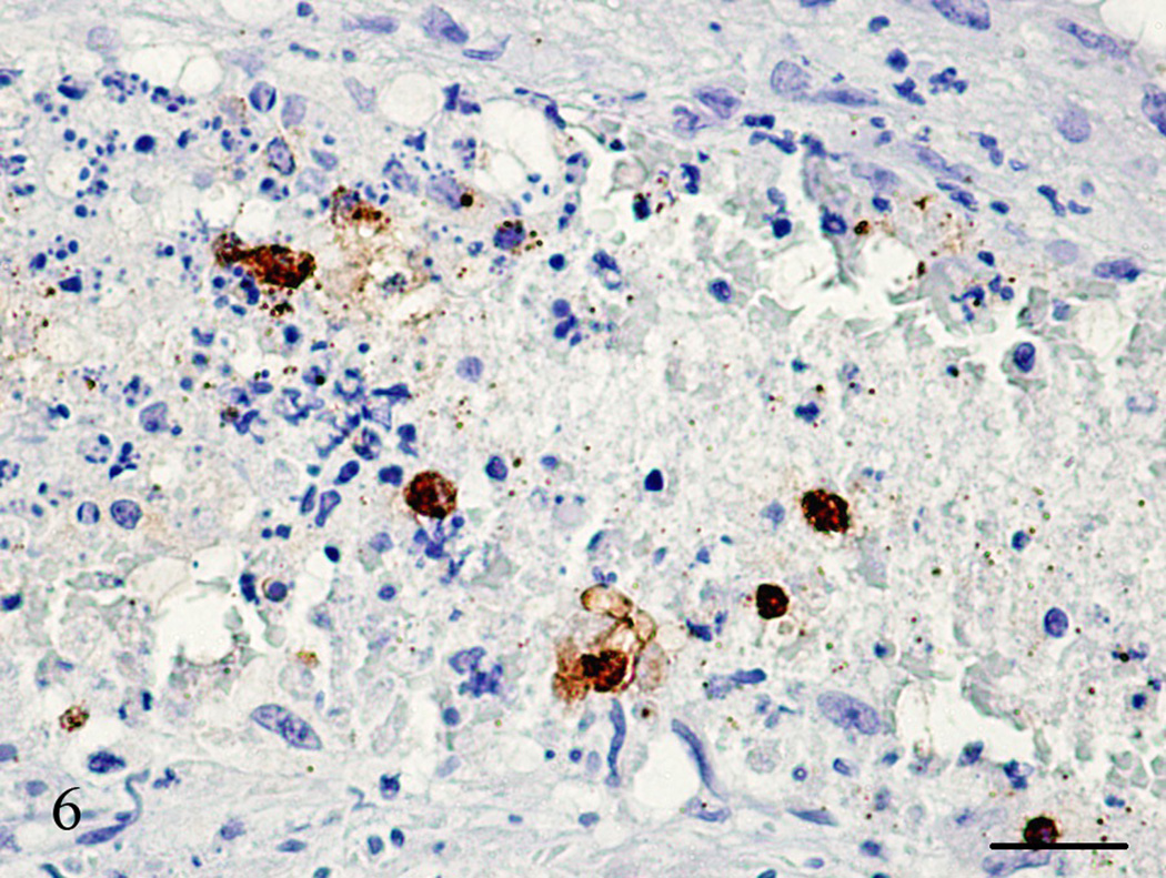
Figure 6.
Lung, Aotus nancymae. Same medium-sized artery lumen from Figure 5 showing an organized thrombus with necrotic cells and a few neoplastic cells demonstrated by being strongly stained with SP-1 Chromogranin. I mmunohistochemistry, peroxidase staining, hematoxylin counter stain. Bar = 50 µm.

