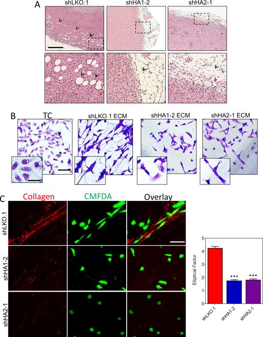
Figure 4.
Collagen prolyl hydroxylase expression promotes invasion. A, representative hematoxylin and eosin staining of the tumor/adipose tissue boundary. Scale bar = 200 μm. B, crystal violet staining of MDA-MB-231 cells plated on tissue culture plastic (TC) or ECM-derived from indicated subclones. Scale bar = 100 μm; inset scale bar = 25 μm. C, CMFDA-labeled naïve MDA-MB-231 cells (green) were seeded on tumor sections and stained with picrosirius red to image collagen fibers (red). Scale bar = 100 μm.

