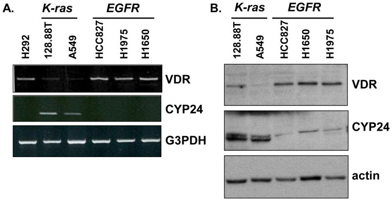
Figure 1. EGFR and K-ras mutation positive NSCLC cells differ in their basal expression of VDR and CYP24.
(A) mRNA expression levels of VDR and CYP24 were evaluated in the indicated cell lines by semi-quantitative RT-PCR. (B) Whole cell extracts were prepared from the indicated cell lines. Equivalent amounts of protein were analyzed by immunoblot for VDR and CYP24. Blots were reprobed for actin as a control for protein quantitation and loading. Results are representative of 3 independent experiments.

