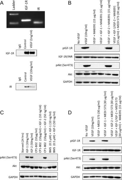
Figure 4.

A. Upper panel: Analysis of IGF-1R and IR, mRNA levels in HUVEC cells by RT-PCR amplification of IGF-1R (389 bp), INS (364 bp). RT-PCR products were analyzed on ethidium bromide-stained agarose gels. Lower panel: Immunoblot of HUVEC extracts demonstrating levels of IGF-1R and IR proteins at time of VEGF stimulation and at 24 hr. B. HUVECs were stimulated with VEGF in the absence or presence of IGF-1 receptor-binding antibody (MAB392; 10 μg/ml; 24 hr) without or with exogenous IGF-1 or IGF-2. C. BMS754807 (0.5 μM) inhibits IGF-1 and IGF-2 stimulated phosphorylation of Akt. HUVECs were starved as above, and incubated for 2 hr with CP1-B02 or BMS754807 prior to stimulation with IGF-1 or IGF-2. D. HUVECs were stimulated with VEGF in the absence of MEDI-573 ligand binding antibody (30 μg/ml; 24 hr) without or with exogenous IGF-1 or IGF-2. Right lane shows stimulation with both ligands in the presence of MAB391 and MEDI-573. Phosphorylation of IGF-1R (Tyr1131) or IR (Tyr1146) and Akt (Ser473) was determined at 24 hr.
