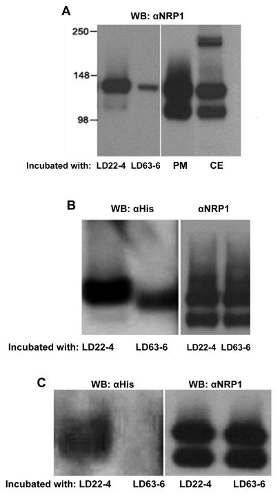
Figure 5.
Co-Immunoprecipitation of LD22-4 and NRP1. A. LD22-4 or LD63-6-Sepharose was incubated with U87MG membrane extracts and the bound protein analyzed for NRP1 by Western blot (left panel). NRP1 in U87MG plasma membrane extracts (PM) or whole cell extracts (CE) were visualized by Western blot (right panel). B. NRP1 was isolated from U87MG membrane extracts by αNRP1-agarose and the agarose-αNRP1-NRP1 complex incubated with LD22-4 or LD63-6. Western blot analysis was performed with α-His antibodies (left panel) to detect LD22-4 and LD63-6. The relative amount of NRP1 presented to LD22-4 and LD63-6 is shown in the right panel. C. LD22-4 or LD63-6 was incubated with intact U87MG cells, the cells extracted, and the extracts incubated with αNRP1-agarose. Agarose complexes were analyzed for LD22-4 and LD63-6 by Western blot. (left panel). The relative amounts of NRP1 captured by the αNRP1-agarose was measured by removing an equal aliquot of agarose after incubation with cell extracts and analyzing them for NRP1 (right panel).

