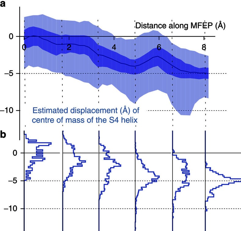
Figure 5. The wild-type PVPV S4 helix is displaced vertically by up to ~8 Å when the pore is operationally closed.
(a) The centre of mass of the S4 helix moves down towards the intracellular side of the lipid bilayer as the kink in the S6 helices is reduced, closing the pore. The average along the minimum free energy path is drawn as a blue line. The ranges 25–50% and 2–98% are shaded medium and light-blue, respectively. (b) The distributions are also plotted for six reference points.

