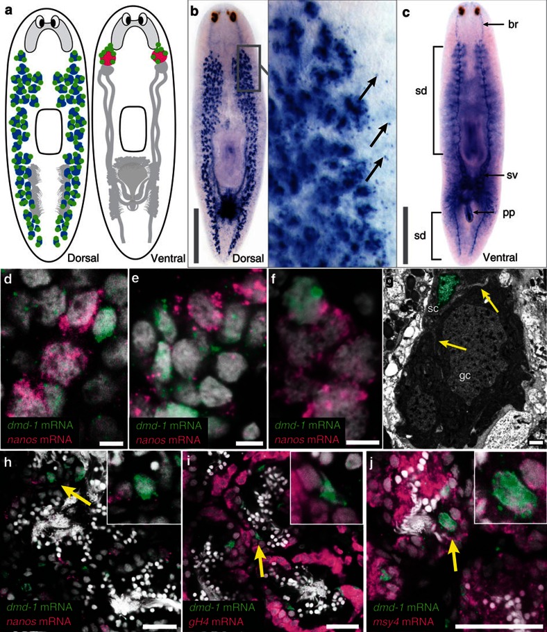
Figure 1. dmd-1 is expressed in the somatic cells of the male reproductive system.
(a) Generalized reproductive system in the sexual planarian. Left, testes (blue) are located dorsolaterally. Right, ovaries (pink) are located more ventrally at base of the brain. nanos-positive germline stem cells of the testes and ovaries (green). Accessory reproductive structures (grey). (b) WISH showing dmd-1 transcripts in and around (arrows) the testes in a sexually mature planarian. (c) Ventral view of the animal showing dmd-1 transcripts in the brain (br), sperm ducts (sd), seminal vesicles (sv) and penis papilla (pp) of a sexually mature planarian. (d–f) Single confocal sections showing two-colour FISH for dmd-1 (green) and nanos (magenta) mRNAs in the testes primordia of animals <24 h after hatching (d), developing testes of sexually immature planarians (e) and germ cell clusters in asexuals (f). (g) TEM showing germ (gc) and somatic cells (sc, nucleus pseudo-coloured green, arrows indicate processes) in an asexual planarian. (h–j) Single confocal sections showing two-colour FISH for dmd-1 (green) and germ cell markers (magenta). Somatic cells expressing dmd-1 in the testes do not express germ cell markers nanos (h, 124/125 cells are dmd-1+/nanos−), gH4 (i, 136/136 cells are dmd-1+/gH4−) and msy4 (j, 129/129 cells are dmd-1+/msy4+). Cell counts were performed from 25–38 testes lobes in at least two animals. Insets, magnified views show cells expressing dmd-1 transcripts (yellow arrows). Scale bars: b,c, 1,000 μm; d–f, 5 μm; g, 1 μm; h–j, 25 μm.

