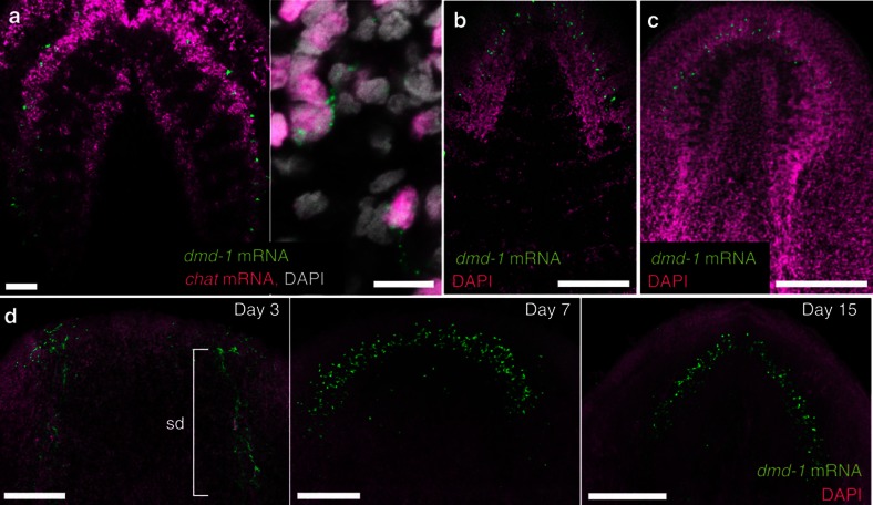
Figure 2. dmd-1 is expressed in some neurons in the planarian brain.
(a) Left, two-colour FISH of dmd-1 and chat transcripts in the brain of a sexual planarian. Right, single confocal section showing cells co-expressing dmd-1 mRNA (green) and chat transcripts (magenta), a marker of cholinergic neurons. (b) dmd-1 transcripts, detected by FISH, in the brain of animals <24 h after hatching. (c) dmd-1 transcripts, detected by FISH, in the brain of an asexual planarian. (d) FISH to detect dmd-1 transcripts in animals during brain regeneration. The sperm ducts (sd) in the old tissue are still present in day 3 regenerates, but are barely visible owing to regression in day 7 and day 15 regenerates. (a–d) Images shown are confocal projections. Scale bars: a, 50 μm; b–d, 200 μm.

