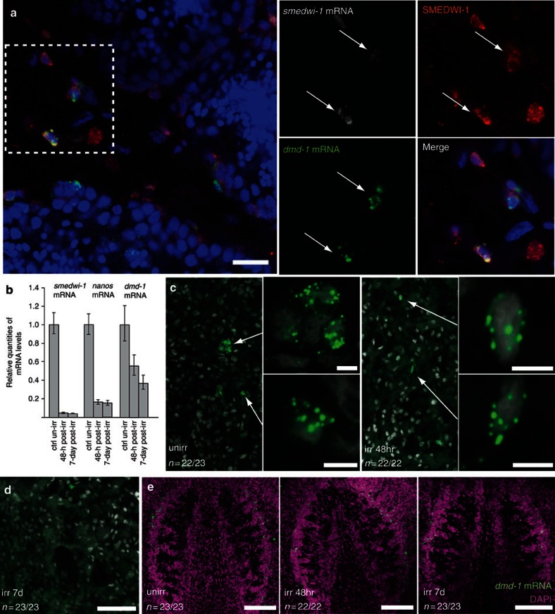
Figure 3. dmd-1-positive cells in proximity to the testes are differentiating neoblast progeny.
(a) Single confocal section showing two-colour FISH combined with immunostaining in mature sexual animals. dmd-1 cells (green) in close proximity to the testes express smedwi-1 transcripts (grey), a marker of neoblasts. SMEDWI-1 protein (red), a marker of neoblasts and differentiating neoblasts is also detected in these dmd-1-positive cells. Arrows indicate dmd-1-positive cells within the dashed box. (b) qPCR showing relative levels of smedwi-1, nanos and dmd-1 mRNAs in irradiated (100 Gy) versus unirradiated asexual planarians 48 h and 7 days post irradiation. Error bars indicate 95% confidence intervals calculated based on s.e.m., results are the mean from three independent experiments using pools of eight planarians. (c) At 48 h after irradiation, dmd-1-positive cells (arrows) are present in the dorsolateral region of the animal, albeit in fewer numbers, compared with control animals. Magnified views of cells expressing dmd-1 transcripts (insets). dmd-1 transcripts were detected by FISH. Images shown are single confocal sections. (d) At 7 days after irradiation, dmd-1-positive cells are rarely detectable in the dorsolateral region of the animal. dmd-1 transcripts were detected by FISH. Images shown are single confocal sections. (e) Confocal projection showing that dmd-1-positive cells (detected by FISH) are still present in the brain at 48 h and 7 days after irradiation. Scale bars: a, 20 μm; c, 5 μm; d, 50 μm; e, 100 μm.

