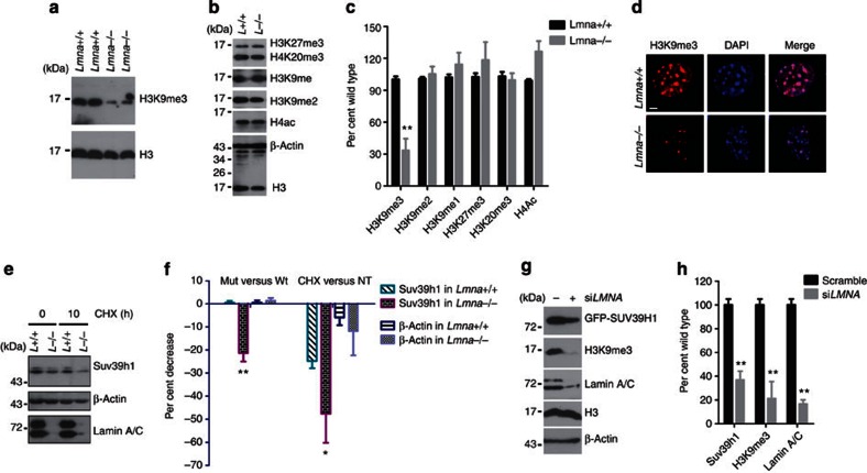
Figure 5. Reduced levels of H3K9me3 and Suv39h1 in Lmna null cells.
(a) Representative immunoblots in MEFs isolated from two pairs of Lmna null and wild-type embryos, showing reduced level of H3K9me3. (b) Representative western blotting showing comparable levels of H3K27me3, H4K20me3, H3K9me1, H3K9me2 and acetyl H4 between Lmna null and wild-type cells. (c) Quantification of experiments in a and b. Data represent mean±s.e.m., n=3. *P<0.05, two-tailed t-test. (d) Representative photos of immunofluorescence staining of H3K9me3 in wild-type and Lmna–/– cells. Scale bar, 5 μm. (e) Representative immunoblots showing expression of Suv39h1 in wild-type and Lmna–/– MEFs treated with CHX (10 μg ml−1). (f) Quantification of experiments in e. Left, per cent changes relative to wild-type cells; right, per cent changes relative to untreated (NT). Data represent mean±s.e.m., n=3. *P<0.05, **P<0.01, two-tailed t-test. (g) Representative western blotting in HEK293 cells stably expressing GFP-SUV39H1 treated with LMNA or scramble siRNA. (h) Quantification of experiments in g. Data represent mean±s.e.m., n=3. **P<0.01, two-tailed t-test.

