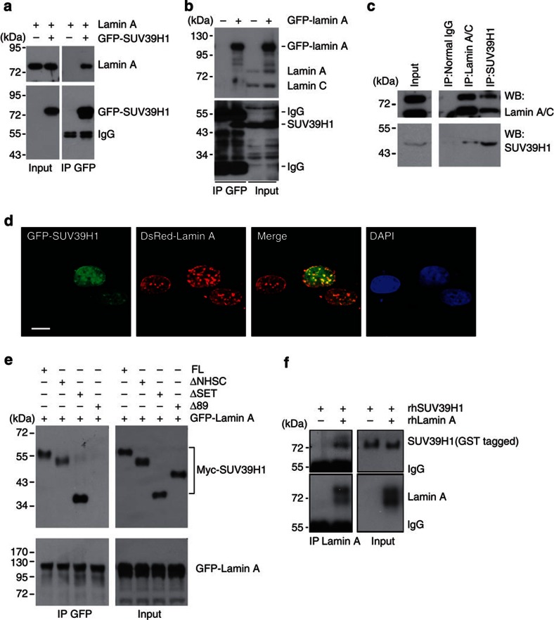
Figure 6. Lamin A interacts with SUV39H1.
(a) GFP-SUV39H1 and/or lamin A were ectopically expressed in HEK293 cells. Representative immunoblots showing ectopic lamin A in the anti-GFP immunoprecipitates. Data are representative of three independent experiments. (b) Representative immunoblots showing endogenous SUV39H1 in the anti-GFP immunoprecipitates in HEK293 cells expressing ectopic GFP-lamin A. (c) Representative western blotting of SUV39H1 and lamin A/C in the anti-SUV39H1 and anti-lamin A/C immunoprecipitates in HEK293 cells. (d) Representative photos of confocal microscopy in human fibroblasts expressing ectopic GFP-SUV39H1 and DsRed-lamin A, showing colocalization of DsRed-lamin A and GFP-SUV39H1 on the nuclear lamina and in the nuclear interior. Scale bar, 10 μm. (e) Representative western blotting showing various Myc-tagged SUV39H1 mutant proteins in the anti-GFP immunoprecipitates in HEK293 cells expressing GFP-lamin A and Myc-tagged SUV39H1 mutants. Note a complete loss of Δ89 in the anti-GFP immunoprecipitates. Abbreviations: FL, full-length SUV39H1; Δ89, N-terminal-89-amino-acid-deleted SUV39H1; ΔSET, SET-domain-deleted SUV39H1; ΔNHSC, NHSC-sequence-deleted SUV39H1. (f) Representative immunoblots showing full-length recombinant human SUV39H1 (rhSUV39H1) in the anti-lamin A immunoprecipitates in the test tube containing recombinant human lamin A (rhLamin A) and rhSUV39H1.

