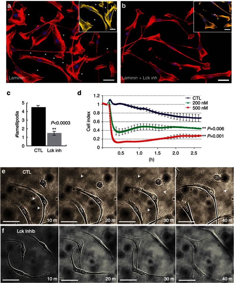
Figure 3. Inhibition of Lck reduces the formation of radial lamellipodia in SCs.
RSCs seeded on a laminin substrate were allowed to attach for 1.5 h, then were left untreated (a) or treated (b) with 500 nM Lck inhibitor for 2 h. Cultures were fixed and stained for phalloidin-rhodamine to identify lamellipodia (asterisks). Insets show magnified images of SCs pseudo-coloured yellow using Photoshop to represent the loss of radial lamellipodia (asterisks) after treatment with Lck inhibitor. (c) Numbers of radial lamellipodia per cell seeded on a laminin substrate were counted (30 cells per well, n=4 from two independent experiments, error bars represent ±s.d.). Treatment with Lck inhibitor significantly reduced the number of radial lamellipodia per cell on a laminin substrate (P<0.0003). (d) RSCs were seeded onto a laminin-coated xCELLigence E-plate and allowed to attach for 2 h. Cell spreading was monitored every 15 s following addition of DMSO (CTL), 200 nM Lck inhibitor or 500 nM Lck inhibitor. Addition of Lck inhibitor induced an immediate retraction of cell processes as compared with DMSO treated cultures (CTL line in graph). A slight increase in cell index, indicating cell spreading, was seen after the acute phase of treatment but overall the cell index remained significantly decreased after 2 h (n=4, P<0.006 (200 nM) and P<0.001 (500 nM) by Student’s t-test, error bars represent ±s.d.). (e,f) RSCs were seeded on laminin substrate and live cell imaging was performed with images acquired every 2 min using a Zeiss Axiovert microscope equipped with the AxioVision Software. Dynamic extension and retraction of radial lamellipodia were seen in control cultures (e, arrowheads) while only initial retraction and no extensions of radial lamellipodia were seen during Lck inhibition (f). Scale bars represent 50 μm.

