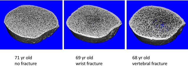
Fig. 5.
HR-pQCT scans of the distal tibia from three women of similar ages in the study that illustrate some of the microarchitectural differences observed between groups, particularly lower trabecular number, increased trabecular separation, and heterogeneity in the fracture subjects compared with the control. These abnormalities were most pronounced in women with vertebral fractures.

