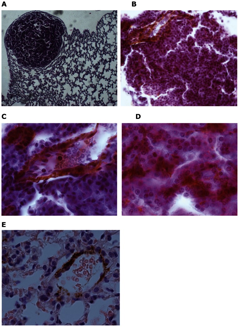
Figure 2.
Histology and immunohistochemistry performed on the lungs of BALB/c mice at 4 months after urethane administration. A representative lung from a urethane-treated mouse showing a nodule after 4 months of follow-up (A, 100x). Global view of a lung nodule showing α-smooth muscle actin expression (B, 400x) and more detail of this expression in a vessel (C, 1000x) and tumor cell (D, 1000x). Lung from a representative control mouse showing α-smooth muscle actin expression in a vessel (E, 1000x).

