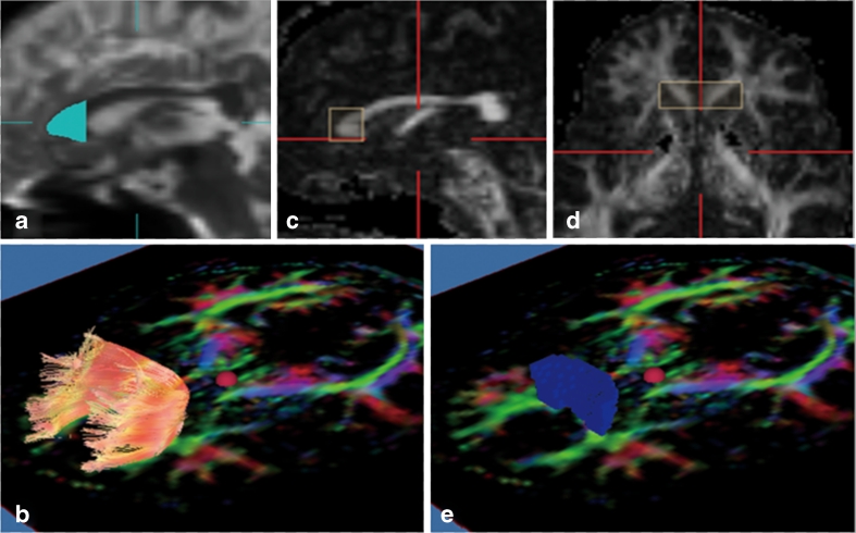
Fig. 1.
Diffusion tensor tractography images of the genu of the corpus callosum and fibre tracts, and voxelisation. a The seed region of interest was placed manually including the entire genu of the corpus callosum (light blue area) on a reconstructed mid-sagittal image with a non-diffusion-weighted image (b = 0 s/mm2). b Tractographic image of the genu of the corpus callosum was generated with threshold values of line-tracking termination as fractional anistropy (FA) > 0.18. c, d Fibre tracts of the genu of the corpus callosum were defined as follows: anterior–posterior, including the first sixth of the corpus callosum; above and below, including the upper and lower border of the genu of the corpus callosum; right–left, between the anterior horns of the lateral ventricles. e Voxelisation was performed along the genu of the corpus callosum (blue voxels). FA and mean diffusivity (MD) values in co-registered voxels were calculated

