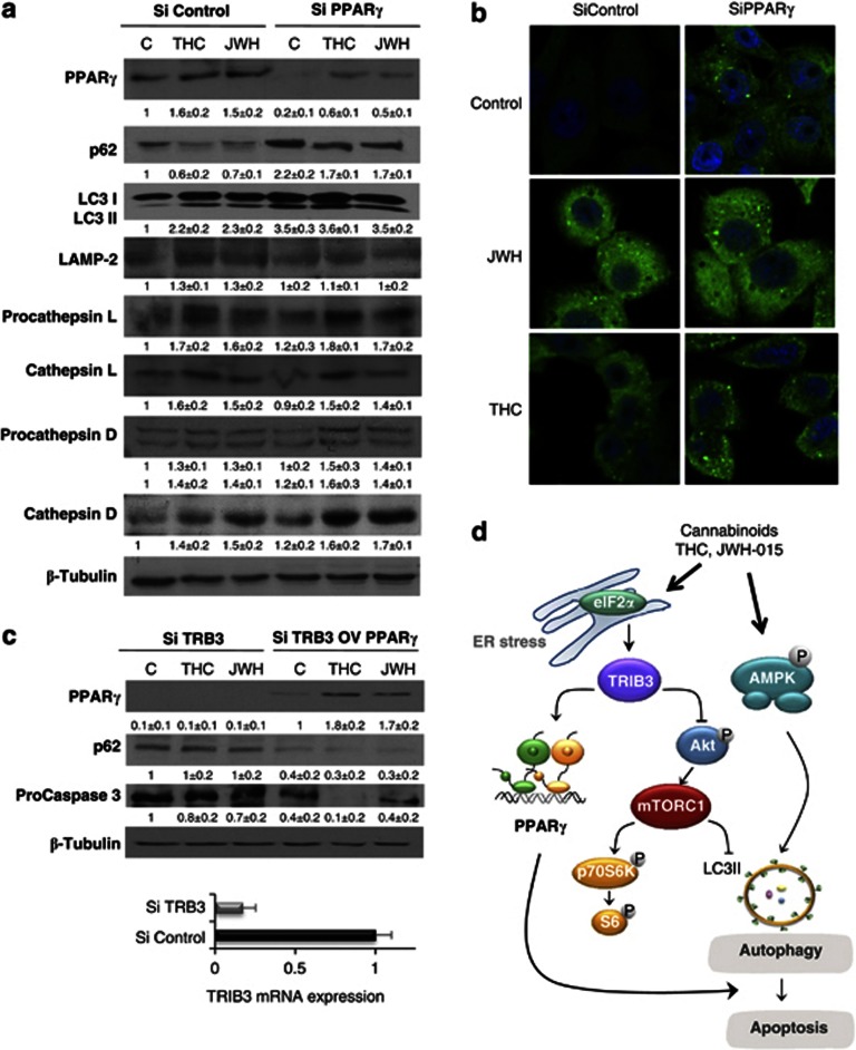
Figure 4.
PPARγ participates in autophagy induction. (a) HepG2 cells were transfected with siRNA control or selective for PPARγ (siControl or siPPARγ) and treated with THC 8 μM or JWH-015 8 μM and PPARγ and p62 levels, microtubule-associated protein 1 light chain 3a non-lipidated (LC3-I) and lipidated (LC3-II) forms, LAMP-2 protein, as well as procathepsin and cathepsil L, procathepsin and cathepsin D, were detected by western blot. Tubulin levels are shown as loading control. A representative western blot of three different experiments is shown. (b) HepG2 cells were transfected with siRNA control or selective for PPARγ (siControl or siPPARγ), treated with THC 8 μM or JWH-015 8 μM for 24 h, and LC3 was detected by confocal immunofluorescence. Nuclei were stained with 4′,6-diamidino-2-phenylindole. (c) HepG2 cells were transfected with scramble (SiControl) or TRIB3 siRNA (SiTRIB3). At the same time, cells were transfected with an empty vector or PPARγ overexpression vector as indicated in the Materials and Methods section. After 48 h, cells were treated with DMSO (control), THC 8 μM or JWH 8 μM for 24 h and PPARγ, p62, procaspase 3 and Tubulin were measured by western blot. Images are representative of three different experiments. Levels of TRIB3 mRNA measured by quantitative PCR in siControl and siTRIB3-treated cells are shown under the western blot. (d) Scheme of the proposed mechanism of cannabinoid-induced HCC cell death. Cannabinoid treatment stimulates autophagy via two different mechanisms: (i) activation of AMPK or (ii) upregulation of tribbles homolog 3. TRIB3 induces subsequent inhibition of the serine–threonine kinase Akt/mammalian target of rapamycin C (Akt/mTORC1) axis or upregulation of PPARγ. Stimulation of autophagy by cannabinoids leads to HCC apoptosis and cell death

