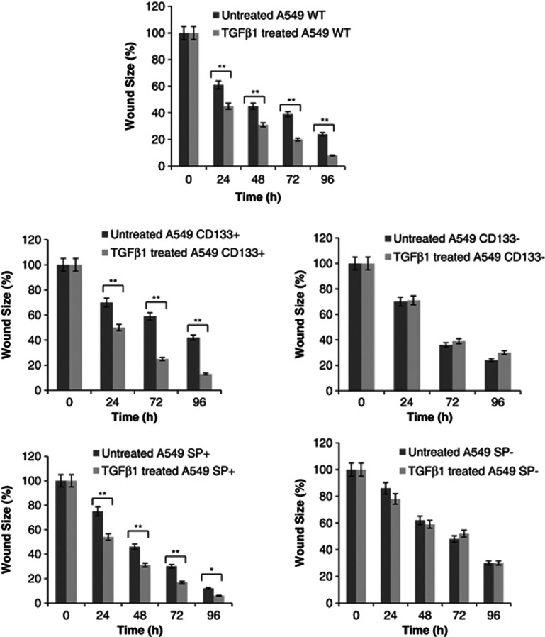
Figure 7.
Wound healing analyses. Wound size analyses performed at 24, 48, 72 and 96 h, after TGF-β1 treatment, showed that TGF-β1 enhanced motility in WT A549 as well as CD133+ and SP+ cells compared to untreated controls. CD133+ cells showed higher motility than other cell fractions. For CD133− and SP− cells, TGF-β1 exposure did not change the motility

