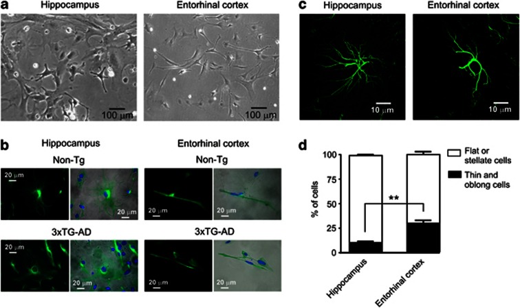
Figure 1.
Morphological differences between cultured astrocytes derived from EC and hippocampus. (a) Microphotographs show the phase-contrast images of live (non fixed) hippocampal cultured astrocytes (left) and EC cultured astrocytes (right) at 4 days in vitro. (b) Representative images of GFAP-stained hippocampal and EC astrocytes derived from non-Tg control and 3xTg-AD model animals. Left panels represent images of GFAP immunofluorescence of stained cells, whereas right panels show merged images of phase-contrast images (gray) and GFAP/DAPI (green/blue) staining obtained from the same cells and fields of view. (c) In situ images of GFAP-stained hippocampal and EC astrocytes; the cells were GFAP labelled in slices obtained from 6-month-old non-Tg animals. For technical details, see Olabarria et al.15 and Yeh et al.22 (d) Quantification of the percentage of thin, long needle-shaped and flat or stellate cells in cultures prepared from hippocampus and the EC; n=320, from four different cultures **P<0.01

