Figure 3.
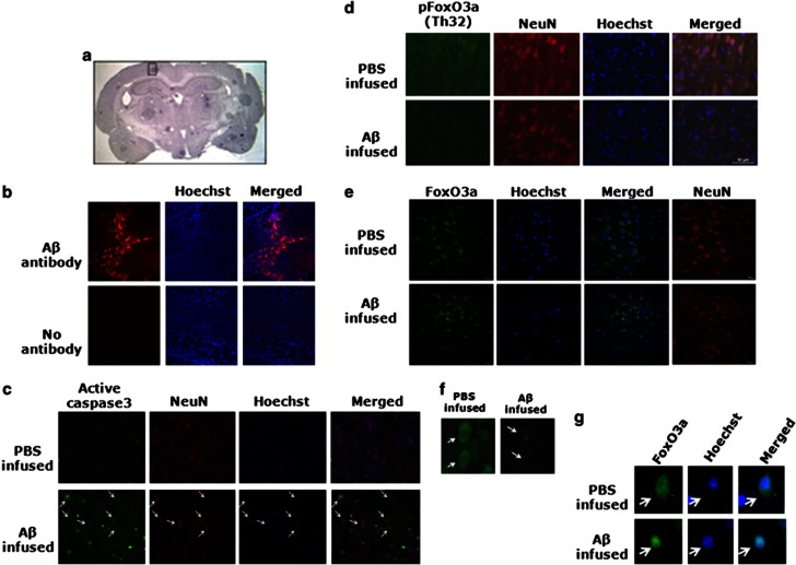
FoxO3a is dephosphorylated following Aβ1-42 infusion in vivo. (a) The rats were infused with either Aβ1-42 or PBS in the right hemisphere of the brain in the treated or control group, respectively. After 21 days of infusion, the animals were killed and the brains were taken out following cardiac perfusion. The frozen brains were sectioned by cryotome. Representative image of a brain section stained with haematoxylin–eosin is shown. (b) Brain sections of the treated rats were immunostained with Aβ1-42 antibody to check the presence of Aβ plaques in the infused area. Upper panel shows brain section stained with anti-Aβ1-42 antibody. Lower panel shows brain sections from the same animal stained without any primary antibody as a negative control. (c) Brain sections (portion marked with black box in a) of control and treated groups were co-immunostained for active caspase3 and NeuN. Nuclei were stained with Hoechst. Arrows show neurons with high active caspase-3 staining. Representative image of six sections from three animals with similar results is shown here. Images were taken under × 40 objective. (d) Brain sections (portion marked with black box in a) of control and treated groups were co-immunostained for p-FoxO3a(Thr32) and NeuN. Nuclei were stained with Hoechst. Representative images of six sections from three animals with similar result are shown here. Scale bar, 50 μm. (e) Confocal images of brain sections from control and treated groups were co-immunostained for FoxO3a and NeuN. Nuclei were stained with Hoechst. Representative images of six sections from three animals with similar results are shown here. Scale bar, 10 μm. (f) Enlarged view of the first column of d, showing cells from treated and control brains immunostained with p-FoxO3a(Th32). Arrows show neurons with high or low p-FoxO3a(Th32) staining. (g) Enlarged view of the first column of e, showing cells from treated and control brains immunostained with FoxO3a. Arrows show the neurons with cytosolic or nuclear FoxO3a

