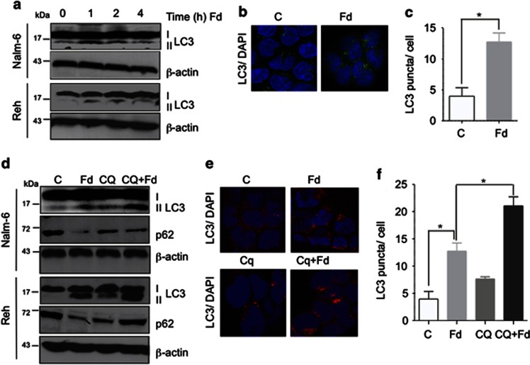
Figure 1.
Fd induces autophagy and enhances autophagic outflux.(a) Western blot for LC3 I/II expression, with β-actin serving as a loading control, in Nalm-6 and Reh cells treated with Fd for the indicated time. (b) Representative images of LC3 stained cytospins of Nalm-6 cells treated with Fd for 1 h. Nuclei were stained with DAPI. Image quantification of LC3 puncta is shown in (c). (d) Western blot for LC3 I/II, p62/SQSTM1(p62) and β-actin as a loading control in Nalm-6 and Reh cells pretreated with CQ for 1 h followed by 4-h treatment with Fd. (e) Confocal immunostaing for LC3-I/II in Nalm-6 cells pretreated with CQ for 1 h followed by 4-h treatment with Fd. Nuclei were stained with DAPI. Quantification of LC3 puncta staining is shown in (f); *P<0.05, n=3

