Figure 5.
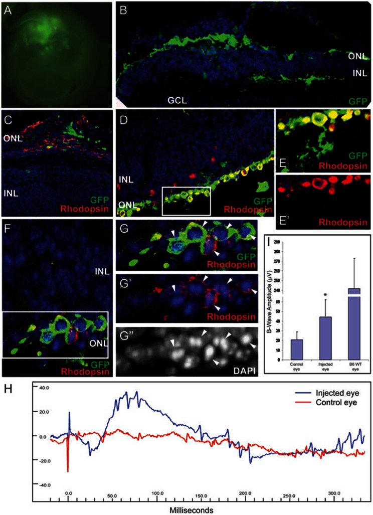
Transplanted photoreceptors from retinal stem cells provide functional and structural retinal rescue in retinitis pigmentosa disease model. (A) Fluorescence microscope image of eye shows large clusters of GFP+ cells 4-5 weeks after FVB/NJ pups are transplanted with photoreceptors derived from retinal stem cells. (B) Histological immunostaining of transplanted retina shows that many transplanted GFP+ cells leave the subretinal space and integrate into the remaining ONL, the OPL and INL and a few cells migrate into IPL layer, while host photoreceptors are degenerated. (C) In some cases, transplanted cells form a new layer between INL and pigmented layer and express rod photoreceptor marker, Rhodopsin. (D–G) Integrated GFP+ cells express Rhodopsin and form condensed nuclei (arrowheads), similar to normal rod photoreceptors. (E–E' and G–G'') are high-power images of the boxed regions in D and F. (H) A representative electroretinograph (ERG) from one mouse. The uninjected control eye (red) has no response to light stimulus, however, the transplanted eye from the same animal has a clear response to a flash of light. (I) Transplanted eyes exhibit significantly increased B-wave amplitudes following light flash compared with control eyes, though not reaching the amplitude exhibited by normal B6 wild-type mouse eyes. Data is shown as mean ± SEM (* P < 0.01). Blue, DAPI.

